페이지

2015년 10월 14일 수요일

2015년 니콘 현미경 사진 수상작: Photographing the Microscopic: Winners of Nikon Small World 2015

Nikon just announced the winners of the 2015 Small World Photomicrography Competition, and they’ve shared some of the winning and honored images from this year’s competition with us here. The contest invites photographers and scientists to submit images of all things visible under a microscope. More than 2,000 entries were received from 83 countries this year. Enjoy a trip into a miniature world through the images below, all from the 2015 Nikon Small World Photomicrography Competition.
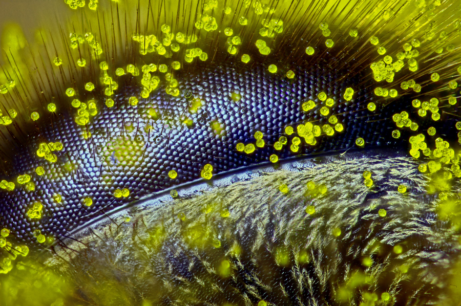
  • First-Place Winner. The eye of a honeybee (Apis mellifera) covered in dandelion pollen (120x). 
    Ralph Claus Grimm, Queensland, Australia
  • 12th Place. Developing sea mullet (Mugil cephalus) embryos (40x). 
    Hannah Sheppard-Brennand, New South Wales, Australia
  • Image of Distinction. The radula, or feeding structure, of a limpet, a kind of aquatic snail (40x). 
    Michael Crutchley, Wales, United Kingdom
  • Image of Distinction. Cross section of a water lily leaf bud (Nupha lutea) (12.5x). 
    Dr. David Maitland, Feltwell, United Kingdom
  • Image of Distinction. Scales on a moth wing (300x). 
    Don Parsons, Wisconsin, US
  • 14th Place. Australian grass (Austrostipa nodosa) seed (5x). 
    Viktor Sykora, Prague, Czech Republic
  • Image of Distinction. Antenna of a male moth (Anisota sp.) (100x). 
    Dr. Igor Siwanowicz, Virginia, USA
  • Honorable Mention. Liverwort (Lepidolaena taylorii) plant showing modified leaves (water sacs), which are often home to aquatic microorganisms such as rotifers (100x). 
    Susan Tremblay, Berkeley, California, USA
  • Image of Distinction. Vascular bundles of papyrus (Cyperus papyrus) (200x). 
    Dr. David Maitland , Feltwell, United Kingdom
  • 19th Place. Planktonic larva of a horseshoe worm (phoronid) (450x). 
    Dr. Richard R. Kirby, Plymouth, United Kingdom
  • Image of Distinction. Rove beetle head (Tychobythinus sp.) (10x). 
    Dr. Joseph Parker, New York, USA
  • Image of Distinction. Colony of single-celled organisms (Carchesium ciliates) (160x). 
    Arturo Agostino, Reggio Calabria, Italy
  • Honorable Mention. Detail of jewel beetle (Coleoptera Buprestidae) (32x). 
    Dr. Luca Toledano, Verona, Italy
  • Image of Distinction. Texture of a chiral thermotropic liquid crystal (20x.) 
    Dr. Giuliano Zanchetta, Milan, Italy
  • Image of Distinction. Vilene fabric (glue drops shown in blue) (80x). 
    Gerd Günther, Düsseldorf, Germany
  • Honorable Mention. Mites on insect pupa (20x). 
    Rogelio Moreno Gill, Panama
  • 20th Place. Suction cups on a diving beetle (Dytiscus sp.) foreleg (50x). 
    Frank Reiser, New York, USA
  • Image of Distinction. Mouth parts (pseudo trachea) of a blowfly (Calliphora vomitoria) (750x). 
    Raymond Morrison Sloss, Oxfordshire, United Kingdom
  • 13th Place. Tentacles of a carnivorous plant (Drosera sp.) (20x). 
    Jose Almodovar, Puerto Rico, USA
  • Image of Distinction. Triple-labeled rat cerebellum (100x). 
    Thomas Deerinck, California, USA
  • Image of Distinction. Jaws and head of a long-jawed spider (Metellina sp.) (10x). 
    Geir Drange, Asker, Norway
  • Image of Distinction. Transverse section of an ostrich fern (250x). 
    Anatoly Mikhaltsov, Omsk, Russia
  • Image of Distinction. Hipparion fossilized bone (100x). 
    Dr. Santiago Gomez, Cadiz, Spain
  • 10th Place. Clam shrimp (Cyzicus mexicanus), live specimen (25x). 
    Ian Gardiner, Alberta, Canada
  • Image of Distinction. Detail of ancient Chinese pottery from the Song Dynasty (960-1126 AD) (4x). 
    Yvonne (Yi-Chieh) Lu, New York, USA
  • 6th Place. Spore capsule of a moss (Bryum sp.). 
    Henri Koskinen, Helsinki, Finland

댓글 없음:

댓글 쓰기